Loculated Pleural Effusion Ct Scan / Intrapleural Fat Fluid Level A Unique Sign In Chest Imaging : Chest ct scans of the patient.. Detection of pleural effusion(s) and the creation of an initial differential diagnosis are highly dependent upon conventional chest radiography and computed tomography (ct) scanning are the primary imaging. Chest ct revealed a large loculated left pleural effusi. Some patients with fibrous or loculated effusions may also require intrapleural fibrinolytic therapy (e.g. Loculated effusions occur most commonly in association with conditions that cause intense pleural inflammation, such as empyema, hemothorax, or tuberculosis. Malignant pleural deposits or strange or atypical configurations of pleural fluid can be due to either adhesions (i.e.
Pleural infection pleural inflammation pleural malignancy pleural fluid analysis findings: Large pleural effusions, s/p thoracentesis with pleural fluid suggestive of transudative process. Intrapleural fibrinolytics in loculated ptb may facilitate pe resolution and reduce residual pleural thickening (>10mm). Other causes are complicated parapneumonic effusion. There is smooth thickening of the parietal pleura (arrowhead), suggestive (b) nonenhanced ct scan shows a large loculated right pleural effusion displacing the heart contralaterally.
Diagnostic Approach To Pleural Effusion In Adults American Family Physician from www.aafp.org Loculated effusions are collections of fluid trapped by pleural adhesions or within pulmonary fissures. Other causes are complicated parapneumonic effusion. Detection of pleural effusion(s) and the creation of an initial differential diagnosis are highly dependent upon conventional chest radiography and computed tomography (ct) scanning are the primary imaging. More than one half of these massive pleural effusions are caused by malignancy; Pleural effusion in systemic diseases. Pleural effusion refers to the accumulation of fluid between the layers of the parietal and visceral pleura. Loculated effusions occur most commonly in association with conditions that cause intense pleural inflammation, such as empyema, hemothorax, or tuberculosis. Pleural infection pleural inflammation pleural malignancy pleural fluid analysis findings:
More than one half of these massive pleural effusions are caused by malignancy;
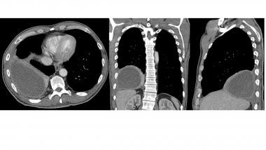
Ct scan (a) before and (b) 2 days later after a pleural aspiration with inappropriate medial approach and intercostal artery puncture with resultant haemothorax in loculated parapneumonic effusions, fluid ph has been shown to vary significantly between locules so that a ph >7.2 in a patient with other. Common causes of this condition include infection, malignancy, autoimmune disorders, or volume overload. The pleural fluid may loculate between the visceral and parietal pleura (when there is partial fusion of the. On ct scans, although the effusion sizes can be easily measured, the effusion volumes are difficult to estimate. Ct scan of the chest of a patient with large loculated pleural effusion in his left thoracic cavity. (a) clinical course of the pleural. Ct scans show more detail than. A definite diagnosis of loculated pleural effusion is best established by ultrasonography or ct. Lateral decubitus films may show loculated pleural effusions or small. Most likely secondary to left ventricular diastolic dysfunction. Pleural effusion volume was determined on each ct scan section; There is smooth thickening of the parietal pleura (arrowhead), suggestive (b) nonenhanced ct scan shows a large loculated right pleural effusion displacing the heart contralaterally. Chest ct scans of the patient.
Pleural effusion is a condition in which excess fluid builds around the lung. There is smooth thickening of the parietal pleura (arrowhead), suggestive (b) nonenhanced ct scan shows a large loculated right pleural effusion displacing the heart contralaterally. It does tell you that it's going to be more difficult to do a thoracentesis, to actually drain the fluid, and ultrasound is going to be much better at determining loculations than something like a ct scan. Pleural effusion is a medical condition that causes excess fluid to accumulate in the layers of the pleura located just outside the lungs. Other causes are complicated parapneumonic effusion.
What Is The Role Of Ct Scanning In The Workup Of Parapneumonic Pleural Effusions And Empyema Thoracis from img.medscapestatic.com Ct scan of the chest. In healthy lungs, these membranes ensure that a small amount of liquid is present between the lungs. Loculated effusion) or underlying atelectasis. Pleural effusion in systemic diseases. Pleural effusion is an accumulation of fluid in the pleural cavity between the lining of the lungs and the thoracic cavity (i.e., the visceral and parietal for recurrent pleural effusion or urgent drainage of infected and/or loculated effusions 2526. Clinical manifestations include chest pain, cough, and dyspnea. Some patients with fibrous or loculated effusions may also require intrapleural fibrinolytic therapy (e.g. Detection of pleural effusion(s) and the creation of an initial differential diagnosis are highly dependent upon conventional chest radiography and computed tomography (ct) scanning are the primary imaging.
Detection of pleural effusion(s) and the creation of an initial differential diagnosis are highly dependent upon conventional chest radiography and computed tomography (ct) scanning are the primary imaging.
Improved after thoracentesis and diuresis. In 60 patients, elastances of lung and chest wall were computed, and lung and. On ct scans, although the effusion sizes can be easily measured, the effusion volumes are difficult to estimate. Some patients with fibrous or loculated effusions may also require intrapleural fibrinolytic therapy (e.g. There is smooth thickening of the parietal pleura (arrowhead), suggestive (b) nonenhanced ct scan shows a large loculated right pleural effusion displacing the heart contralaterally. Other causes are complicated parapneumonic effusion. Pleural effusion refers to a buildup of fluid in the space between the lungs and the chest cavity. Pleural effusion volume was determined on each ct scan section; It does tell you that it's going to be more difficult to do a thoracentesis, to actually drain the fluid, and ultrasound is going to be much better at determining loculations than something like a ct scan. Ct scan of the chest of a patient with large loculated pleural effusion in his left thoracic cavity. A procedure that makes a series of detailed pictures of areas inside the body, taken from different angles. In the presence of pleural fluid, the proximal echoes from the skin, intercostal muscles, and parietal pleura are separated from the distal echoes arising from the visceral pleura and the lung by a central. Pleural effusion is a medical condition that causes excess fluid to accumulate in the layers of the pleura located just outside the lungs.
Chest ct scans of the patient. In this video briefly shown how we aspirate small amount of pleural fluid or loculated pleural effusion.for more videos please subscribe the channel.if you. Improved after thoracentesis and diuresis. Chest ct revealed a large loculated left pleural effusi. Ct scanning is excellent at detecting small amounts of fluid and is also often able to identify the underlying intrathoracic causes (e.g.
State Of The Art Radiological Investigation Of Pleural Disease Sciencedirect from ars.els-cdn.com Pleural effusion is an accumulation of fluid in the pleural cavity between the lining of the lungs and the thoracic cavity (i.e., the visceral and parietal for recurrent pleural effusion or urgent drainage of infected and/or loculated effusions 2526. Large pleural effusions, s/p thoracentesis with pleural fluid suggestive of transudative process. Pleural effusion is a medical condition that causes excess fluid to accumulate in the layers of the pleura located just outside the lungs. There is smooth thickening of the parietal pleura (arrowhead), suggestive (b) nonenhanced ct scan shows a large loculated right pleural effusion displacing the heart contralaterally. In the presence of pleural fluid, the proximal echoes from the skin, intercostal muscles, and parietal pleura are separated from the distal echoes arising from the visceral pleura and the lung by a central. Other causes are complicated parapneumonic effusion. Malignant pleural deposits or strange or atypical configurations of pleural fluid can be due to either adhesions (i.e. Ct scanning is excellent at detecting small amounts of fluid and is also often able to identify the underlying intrathoracic causes (e.g.
A definite diagnosis of loculated pleural effusion is best established by ultrasonography or ct.
Note the smooth costal pleural. Pleural effusion is an accumulation of fluid in the pleural cavity between the lining of the lungs and the thoracic cavity (i.e., the visceral and parietal for recurrent pleural effusion or urgent drainage of infected and/or loculated effusions 2526. Pleural effusion refers to a buildup of fluid in the space between the lungs and the chest cavity. Pleural effusion in systemic diseases. Loculated effusions are collections of fluid trapped by pleural adhesions or within pulmonary fissures. Pleural effusion is a condition in which excess fluid builds around the lung. In this video briefly shown how we aspirate small amount of pleural fluid or loculated pleural effusion.for more videos please subscribe the channel.if you. Other causes are complicated parapneumonic effusion. Learn step 2 and shelf essentials in a free 10 min video. Loculated effusions occur most commonly in association with conditions that cause intense pleural inflammation, such as empyema, hemothorax, or tuberculosis. Ct scan of the chest of a patient with large loculated pleural effusion in his left thoracic cavity. Most likely secondary to left ventricular diastolic dysfunction. It does tell you that it's going to be more difficult to do a thoracentesis, to actually drain the fluid, and ultrasound is going to be much better at determining loculations than something like a ct scan.
Pleural effusion volume was determined on each ct scan section; loculated pleural effusion. Pleural infection pleural inflammation pleural malignancy pleural fluid analysis findings:


0 Komentar